Tetralogy of Fallot
Synonyms :
- TDF
Description
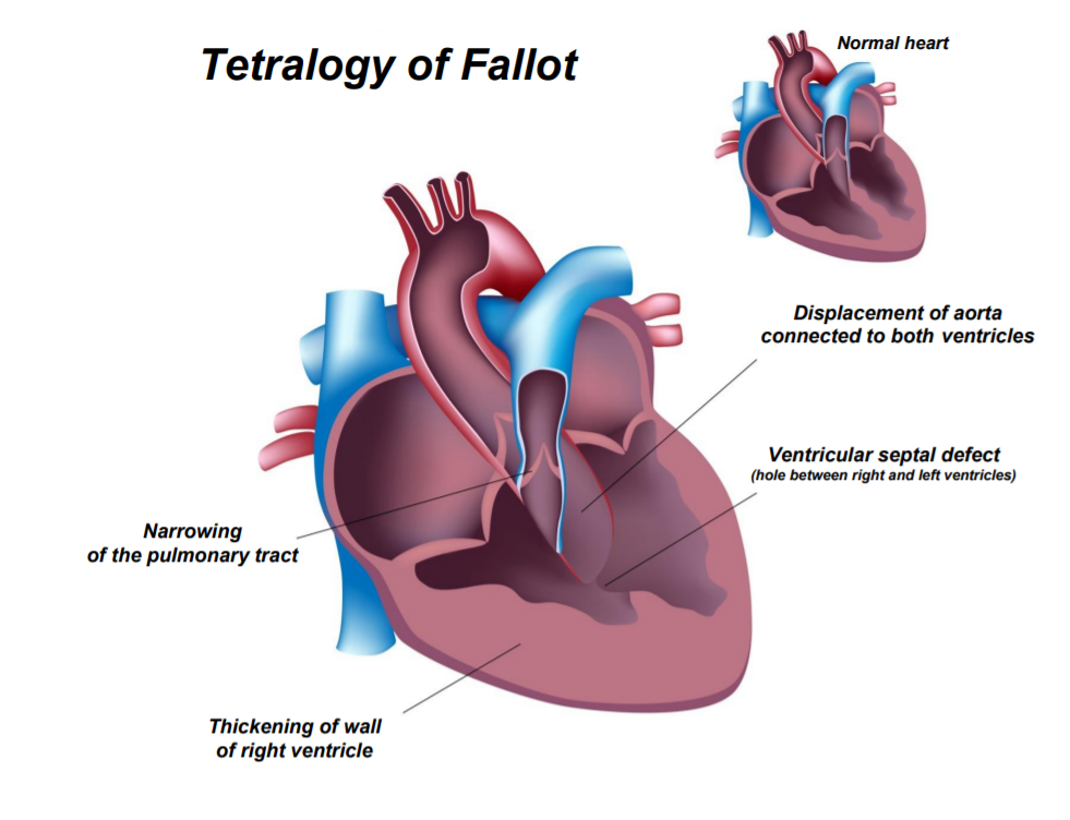
Tetralogy of Fallot is secondary to an anterior deviation of the upper part of the interventricular septum (septum conal).
This deviation is responsible for interventricular communication, aortic overlap on the interventricular septum (dextroposition of the aorta or riding aorta), and sub-pulmonary obstruction (pulmonary stenosis). Besides that, there is a right ventricular hypertrophy.

Incidence
1/2500 - 1/4000
and it represents 10% of congenital heart disease
Codification :
OMIM : 187500 & 618780
CIM-10 : Q21.3
HPO : 0001636
A little story
Its name is associated with Etienne-Louis Arthur Fallot who described it in 1888.
The term "tetralogy of Fallot" has only been used since 1924.
Redo your diagnosis
With Sonio Diagnostics, artificial intelligence that optimises your antenatal diagnosis.
During a quick appointment, share with us the signs you have spotted on the ultrasound, we will replay the exam in Sonio Diagnostics. These appointments are reserved for healthcare professionals.
Ultrasound diagnosis
Ultrasound signs detectable in the 1st trimester
- four-chamber view :
cardiac axis deviation to the left
perimembranous ventricular septal defect - three vessels and trachea view :
discrepant size between the aorta and pulmonary artery with antegrade flow in both vessels on color Doppler - left ventricular outflow tract view :
overriding of the dilated aorta over the ventricular septal defect
ultrasound signs detectable in the 2nd & 3rd trimester
- four-chamber view :
interventricular communication
right ventricular hypertrophy (rarely present in the antenatal period) - left ventricular outflow tract view :
the ventricular septum with the anterior wall of the aorta is lost
aortic overlap on the interventricular septum
interventricular communication - right ventricular outflow tract view :
pulmonary stenosis
discrepant size between the aorta and pulmonary artery - three vessels and trachea view :
discrepant size between the aorta and pulmonary artery
Reversal of flow in arterial duct if pulmonary severe obstruction
Differential Diagnosis
The other conotruncal anomalies that share the same ultrasound call sign of the "aortic overlap on interventricular communication" are :
- isolated sub-aortic interventricular communication where the pulmonary pathway is of normal caliber
- pulmonary atresia with open septum (PAOS) where, on the contrary, the pulmonary ring is atretic, impermeable, and the pulmonary trunk so hypoplastic that it is usually not visible on ultrasound. Of the pulmonary route, only the bifurcation and the pulmonary arteries remain, supplied against the current by the arterial duct.
- the common arterial trunk malformation where, a single artery (the "common trunk") gives rise to both the aorta and the pulmonary arteries.
and also : - transposition of the great arteries
Associated anomalies
Nuchal translucency with normal karyotype
Right aortic arch
Association with extracardiac anomalies is frequent : in particular gastrointestinal and thoracic ones (esophageal and duodenal atresia, and diaphragmatic hernia), even independently of chromosomal anomalies.
Thymic hypoplasia, polyhydramnios.
Risk of chromosomal anomalies
High : 20%
Trisomy 13, 18, 21
Trisomy 8p
del 22q11.2 (Di George syndrome)
Risk of nonchromosomal syndromes
Relatively high
CHARGE syndrome, VACTERL, Fetal alcohol syndrome, Goldenhar syndrome, 4p syndrome, Cornelia Delange syndrome, TAR syndrome, Nager syndrome, Alagille syndrome, Noonan syndrome, Kabuki syndrome, Melnick-Needles syndrome, Townes-Brocks syndrome, 3C syndrome, Coffin-Siris syndrome, WAGR syndrome, , Holt-Oram Syndrome, Yunis-Varon syndrome, Steinfeld syndrome, McKusick Kaufman syndrome
Etiologies - others

With Sonio Diagnostics
Decision support software for prenatal diagnosis, resulting from 5 years of research and collaboration between leading experts in fetal medicine and artificial intelligence, you can combine multiple types of risk factors and ultrasound signs to optimise your diagnostic performance and reduce the risk of errors on almost 300 antenatal diagnosable syndromes* in real time.
Sonio Diagnostics
is CE marked and reserved for fetal ultrasound professionals, and complements their expertise

Some key figures
Recognisable syndromes
Recognisable signs
Prenatal diagnosis practitioners have already adopted it

References
- Lev M, Eckner FA. The pathologic anatomy of tetralogy of Fallot and its variants. Disease Chest 1964; 45: 251–61.
- Hoffman JI, Christianson R. Congenital heart disease in a cohort of 19,502 births with long-term follow-up. Am J Cardiol. 1978;42:641–647.
- Baker EJ. Tetralogy of Fallot with pulmonary atresia. In: Anderson RH, Macartney FJ, Shinebourne EA, et al. eds. Paediatric Cardiology. Edinburgh: Churchill Livingstone, 2001; 1251–79.
- Dupont C, Grati FR, Choy KW, et al. Prenatal diagnosis of 24 cases of microduplication 22q11.2 : an investigation of phenotype-genotype correlations. Prenat Diagn 2015 ; 35(1) : 35–43.
- Boudjemline Y, Fermont L, Le Bidois J, et al. Prevalence of 22q11 deletion in fetuses with conotruncal cardiac defects: a 6-year prospective study. J Pediatr. 2001;138:520–524.
- Rauch R, Hofbeck M, Zweier C, et al.: Comprehensive genotypephenotype analysis in 230 patients with tetralogy of Fallot. J Med Genet 47:321, 2010.
- Goldmuntz E, Geiger E, Benson DW: NKX2.5 mutations in patients with tetralogy of Fallot. Circulation 104:2565, 2001.
- De Luca A, Sarkozy A, Ferese R, et al.: New mutations in ZFPM2/FOG2 gene in tetralogy of Fallot and double outlet right ventricle. Clin Genet 80:184, 2011.
- Hoffman JI, Christianson R. Congenital heart disease in a cohort of 19,502 births with long-term follow-up. Am J Cardiol. 1978;42:641–647.
- Poon LCY, Huggon IC, Zidere V, et al. Tetralogy of Fallot in the fetus in the current era. Ultrasound Obstet Gynecol. 2007;29:625–627.
- Chaoui R, Kalache KD, Heling KS, et al. Absent or hypoplastic thymus on ultrasound: a marker for deletion 22q11.2 in fetal cardiac defects. Ultrasound Obstet Gynecol. 2002;20:546–552.
- Chaoui R, Heling KS, Sarut Lopez A, et al. The thymic-thoracic ratio in fetal heart defects: a simple way to identify fetuses at high risk for microdeletion 22q11. Ultrasound Obstet Gynecol. 2011;37:397–403.
- Zidere V, Tsapakis EG, Huggon IC, Allan LD. Right aortic arch in the fetus. Ultrasound Obstet Gynecol. 2006;28:876–81.
- Pepas LP, Savis A, Jones A, Sharland GK, Tulloh RMR, Simpson JM. An echocardiographic study of tetralogy of Fallot in the fetus and infant. Cardiol Young. 2003;13:240–7.
- Quartermain MD, Glatz AC, Goldberg DJ, Cohen MS, Elias MD, Tian Z, Rychik J. Pulmonary outflow tract obstruction in fetuses with complex congenital heart disease: predicting the need for neonatal intervention. Ultrasound Obstet Gynecol. 2013;41:47–53.
- Vesel S, Rollings S, Jones A, Callaghan N, Simpson J, Sharland GK. Prenatally diagnosed pulmonary atresia with ventricular septal defect: echocardiography, genetics, associated anomalies and outcome.
Heart. 2006;92:1501–5. - Vigneswaran TV, Homfray T, Allan LD, Simpson JM, Zidere V. Persistently elevated nuchal translucency and the fetal heart. J Matern Neonatal Med. 2017:1–5.
- Emami-Moghaddam A, Barati M, Amirpour R, Shojaei K. Prenatal and postnatal echocardiography in NT fetuses with normal karyotype. J Family Med Prim Care. 2019 Aug 28;8(8):2667-2670.
- Vogel M, Sharland GK, McElhinney DB, Zidere V, Simpson JM, Miller OI, Allan LD. Prevalence of increased nuchal translucency in fetuses with congenital cardiac disease and a normal karyotype. Cardiol Young. 2009 Sep;19(5):441-5.
- Hornberger LK, Sanders SP, Sahn DJ, et al. In utero pulmonary artery and aortic growth and potential for progression of pulmonary outflow tract obstruction in tetralogy of Fallot. J Am Coll Cardiol 1995; 25: 739–45.
- Escribano D, Herraiz I, Granados M, et al. Tetralogy of Fallot: prediction of outcome in the mid-second trimester of pregnancy. Prenat Diagn 2011; 31: 1126–33.
- Paladini D, Rustico MA, Todros T, et al. Conotruncal anomalies in prenatal life. Ultrasound Obstet Gynecol 1996; 8: 241–6.

